Under the magnifying eye of a microscope, even an ordinary windowsill can reveal a world of miniature marvels.
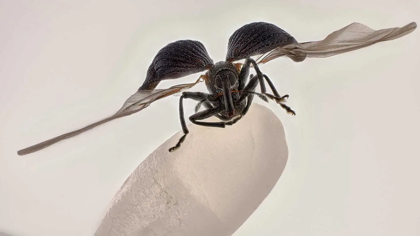
The top honor in the 2025 Nikon Small World Photomicrography Competition went to an image of a rice weevil caught mid–wing stretch atop a single grain of rice. The photo, taken by Zhang You of the Entomological Society of China, was born from a moment of pure chance — he discovered the tiny insect while tidying his home. To capture its delicate form in “flight,” Zhang stacked more than 100 high-resolution microscope shots, creating a portrait that fuses art and science.
The rice weevil (Sitophilus oryzae) is a notorious pest that consumes stored grains and seeds. Zhang had seen infestations before, but finding a naturally preserved specimen with its wings outspread was a rare stroke of luck. “It’s extremely difficult to manually prepare spread-wing samples at this scale,” he explained. “This one was beautifully intact.”
To emphasize the insect’s minuscule size, Zhang mounted it on a single grain of rice. A first-time winner, he said he hopes the image not only showcases scientific detail but also celebrates the elegance of nature’s small designs. “The best work blends artistry with precision,” Zhang said. “It captures the energy and spirit of these tiny creatures.”
Zhang’s winning photo was one of 71 images recognized on October 15 in this year’s competition. Here are a few of the other standout entries.
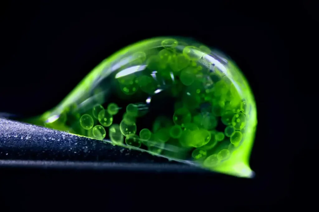
A Drop Teeming with Life
A droplet of water balanced on a syringe tip brims with green algae cells — a microscopic universe in motion.
German chemist and photographer Jan Rosenboom used a 50-year-old microscope he first bought as a teenager, illuminating the scene with LED lights. Inside the droplet, colonies of Volvox algae form swirling spheres — larger green outlines encase smaller, brighter dots representing the next generation of cells developing within.
The droplet perches delicately at an angle on the syringe, offering a perfect sense of scale. Rosenboom admits he spilled over 20 drops before capturing the image that won second place.
He hopes it inspires curiosity about unseen biodiversity: “Even the smallest drop of water can hold entire ecosystems worth protecting.”
Fluorescent Ferns
Igor Siwanowicz, a researcher at the Howard Hughes Medical Institute’s Janelia Research Campus in Virginia, took fifth place with a glowing cross-section of fern spores.
The vivid image, captured using a confocal microscope and fluorescent lasers, reveals three podlike structures — sporangia — where spores develop. The left pod shows brainlike shapes of maturing spores, the middle remains sealed, and the right is empty, likely having burst during preparation. The surrounding frond tissue shines in fiery orange.
“Microscopy can look alien,” Siwanowicz said. “That strangeness sparks curiosity — it makes people want to understand what they’re seeing.”
Sprawling Neurons
At first glance, Stella Whittaker’s image could be mistaken for a human iris. In reality, it depicts the intricate architecture of sensory neurons.
Then a biophysicist at the National Institute of Neurological Disorders and Stroke, Whittaker used fluorescence and antibodies to color-code the sample: actin appears as pink specks, microtubules glow blue and green, and the dark center houses about 10,000 cells.
These delicate neuron branches — found in the limbs and spinal cord — help humans perceive touch. Now pursuing her Ph.D. at Weill Cornell, Whittaker says studying their structure could shed light on how diseases like ALS and Alzheimer’s damage cellular “transport highways.”
Hidden Beauty in Sunflower Hairs
Instead of placing sunflowers in a vase, Polish photographer Marek Miś sliced one under his microscope. The result: a field of ghostly, glowing trichomes — tiny, hairlike structures that protect the plant.
Using polarized light filters of his own design, Miś stacked over 100 images to reveal the trichomes’ extraordinary variation in size and shape. The resulting photo, alive with blues and magentas, shows what the naked eye cannot.
“Sunflowers are already beautiful,” he said. “But under the microscope, they reveal another layer of hidden beauty.”
The Glamour of the Gut
In a striking image from Switzerland’s Friedrich Miescher Institute for Biomedical Research, biologist Marius Mählen and his colleagues turned a mouse colon into a kaleidoscope of color.
Over a single weekend, the team captured 600 confocal microscope images and stitched them together to create the final composite. Antibody staining added contrast: cell nuclei appear white, membranes red, and a cluster of immune cells glows near the left side of the frame.
By studying how healthy tissue forms, Mählen’s team hopes to better understand why cancer and inflammatory diseases create abnormal growths. “If we can learn the rules of healthy development,” he said, “we can start to see how those rules break.”